ohai.social is one of the many independent Mastodon servers you can use to participate in the fediverse.
A cozy, fast and secure Mastodon server where everyone is welcome. Run by the folks at ohai.is.

Administered by:

Server stats:

1.8K
active users

#cytoskeletal

0 posts0 participants0 posts today

📰 "Theory of multiscale epithelial mechanics under stretch: from active gels to vertex models"
biorxiv.org/content/10.1101/20 #Cytoskeletal #Mechanical #Mechanics

bioRxiv · Theory of multiscale epithelial mechanics under stretch: from active gels to vertex modelsEpithelial monolayers perform a variety of mechanical functions, which include maintaining a cohesive barrier or developing 3D shapes, while undergoing stretches over a wide range of magnitudes and loading rates. To perform these functions, they rely on a hierarchical organization, which spans molecules, cytoskeletal networks, adhesion complexes and junctional networks up to the tissue scale. While the molecular understanding and ability to manipulate cytoskeletal components within cells is rapidly increasing, how these components integrate to control tissue mechanics is far less understood, partly due to the disconnect between theoretical models of sub-cellular dynamics and those at a tissue scale. To fill this gap, here we propose a formalism bridging active-gel models of the actomyosin cortex and 3D vertex-like models at a tissue scale. We show that this unified framework recapitulates a number of seemingly disconnected epithelial time-dependent phenomenologies, including stress relaxation following stretch/unstretch maneuvers, active flattening after buckling, or nonreciprocal and non-affine pulsatile contractions. We further analyze tissue dynamics probed by a novel experimental setup operating in a pressure-controlled ensemble. Overall, the proposed framework systematically connects sub-cellular cortical dynamics and tissue mechanics, and ties a variety of epithelial phenomenologies to a common sub-cellular origin. ### Competing Interest Statement The authors have declared no competing interest.

📰 "Time irreversibility, entropy production and effective temperature are independently regulated in the actin cortex of living cells"
arxiv.org/abs/2503.17016 #Physics.Bio-Ph #Cytoskeletal #Dynamics #Actin

arXiv logo
arXiv.orgTime irreversibility, entropy production and effective temperature are independently regulated in the actin cortex of living cellsLiving cells exhibit non-equilibrium dynamics emergent from the intricate interplay between molecular motor activity and its viscoelastic cytoskeletal matrix. The deviation from thermal equilibrium can be quantified through frequency-dependent effective temperature or time-reversal symmetry breaking quantified e.g. through the Kullback-Leibler divergence. Here, we investigate the fluctuations of an AFM tip embedded within the active cortex of mitotic human cells with and without perturbations that reduce cortex activity through inhibition of material turnover or motor proteins. While inhibition of motor activity significantly reduces both effective temperature and time irreversibility, inhibited material turnover leaves the effective temperature largely unchanged but lowers the time irreversibility and entropy production rate. Our experimental findings in combination with a minimal model highlight that time irreversibility, effective temperature and entropy production rate can follow opposite trends in active living systems, challenging in particular the validity of effective temperature as a proxy for the distance from thermal equilibrium. Furthermore, we propose that the strength of thermal noise and the occurrence of time-asymmetric deflection spikes in the dynamics of regulated observables are inherently coupled in living systems, revealing a previously unrecognized link between entropy production and time irreversibility.

📰 "Axonal Mechanotransduction Drives Cytoskeletal Responses to Physiological Mechanical Forces"
doi.org/doi:10.1101/2025.02.11
pubmed.ncbi.nlm.nih.gov/399904
#Mechanotransduction #Cytoskeletal #Mechanical

bioRxiv · Axonal Mechanotransduction Drives Cytoskeletal Responses to Physiological Mechanical ForcesAxons experience strong mechanical forces due to animal movement. While these forces serve as sensory cues in mechanosensory neurons, their impact on other neuron types remains poorly defined. Here, we uncover signaling that controls an axonal cytoskeletal response to external physiological forces and plays a key role in axonal integrity. Live imaging of microtubules at single-polymer resolution in a C. elegans motor neuron reveals local oscillatory movements that fine-tune polymer positioning. Combining cell-specific chemogenetic silencing with targeted degradation alleles to distinguish neuron-intrinsic from extrinsic regulators of these movements, we find that they are driven by muscle contractions and require the mechanosensitive protein Talin, the small GTPase RhoA, and actomyosin activity in the axon. Genetic perturbation of the axon’s ability to buffer tension by disrupting the spectrin-based membrane-associated skeleton leads to RhoA hyperactivation, actomyosin relocalization to foci at microtubule ends, and converts local oscillations into processive bidirectional movements. This results in large gaps between microtubules, disrupting coverage of the axon and leading to its breakage and degeneration. Notably, hyperpolarizing muscle or degrading components of the mechanotransduction signaling pathway in the axon rescues cytoskeletal defects in spectrin-deficient axons. These results identify mechanisms of an axonal cytoskeletal response to physiological forces and highlight the importance of force-buffering and mechanotransduction signaling for axonal integrity. ### Competing Interest Statement The authors have declared no competing interest.

📰 "From Cell Architecture to Mitochondrial Signaling: Role of Intermediate Filaments in Health, Aging, and Disease"
doi.org/doi:10.3390/ijms260311
pubmed.ncbi.nlm.nih.gov/399408
#Cytoskeletal #Mechanical #Cell

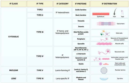
MDPIFrom Cell Architecture to Mitochondrial Signaling: Role of Intermediate Filaments in Health, Aging, and DiseaseThe coordination of cytoskeletal proteins shapes cell architectures and functions. Age-related changes in cellular mechanical properties have been linked to decreased cellular and tissue dysfunction. Studies have also found a relationship between mitochondrial function and the cytoskeleton. Cytoskeleton inhibitors impact mitochondrial quality and function, including motility and morphology, membrane potential, and respiration. The regulatory properties of the cytoskeleton on mitochondrial functions are involved in the pathogenesis of several diseases. Disassembly of the axon’s cytoskeleton and the release of neurofilament fragments have been documented during neurodegeneration. However, these changes can also be related to mitochondrial impairments, spanning from reduced mitochondrial quality to altered bioenergetics. Herein, we discuss recent research highlighting some of the pathophysiological roles of cytoskeleton disassembly in aging, neurodegeneration, and neuromuscular diseases, with a focus on studies that explored the relationship between intermediate filaments and mitochondrial signaling as relevant contributors to cellular health and disease.

📰 "More ATP Does Not Equal More Contractility: Power And Remodelling In Reconstituted Actomyosin"
arxiv.org/abs/2108.00764 #Physics.Bio-Ph #Cond-Mat.Soft #Cytoskeletal #Mechanical #Actomyosin

arXiv.orgMore ATP Does Not Equal More Contractility: Power And Remodelling In Reconstituted ActomyosinThe cytoskeletal component actomyosin is a canonical example of active matter since the powerstroke cycle locally converts chemical energy in the form of adenoside triphosphate (ATP) into mechanical work for remodelling. Observing myosin II minifilaments as they remodel actin {\it in vitro}, we now report that: at high concentrations of ATP, myosin minifilaments form metastable swirling patterns that are characterised by recurrent vortex and spiral-like motifs, whereas; at low concentrations of ATP, such structures give way to aster-like patterns. To explain this, we construct the (quasi-)steady states of a polar active hydrodynamic theory of actomyosin whose ATP-scaling is obtained from a microscopic, stochastic description for the ATP-dependent binding of the heads of single myosin II minifilaments. The latter codifies the heuristic that, since the powerstroke cycle involves the unbinding of myosin II heads from actin, increases in the concentration of ATP reduce the likelihood that a given myosin II minifilament has more than one head bound simultaneously, reducing its ability to generate contractile forces and increasing the relative likelihood of processive motion. This reproduces several qualitative and some quantitative aspects of experiments, providing evidence for the central phenomenon of the theory: an ATP-dependent active contractile instability. ATP therefore controls not only the rate at which work is done -- \textit{i.e.,} the power -- but also the mode by which this occurs.

"We show that migrating #neurons in mice possess a growth cone at the tip of their leading process, similar to that of #axons, in terms of the #cytoskeletal dynamics and functional responsivity through protein tyrosine #phosphatase receptor type sigma (PTPσ). Migrating-neuron growth cones respond to chondroitin sulfate (CS) through PTPσ and collapse, which leads to inhibition of #neuronal migration."

nature.com/articles/s41467-024

NatureIdentification of the growth cone as a probe and driver of neuronal migration in the injured brain - Nature CommunicationsStructure and functions of the tip of migratory neurons remain elusive. Here, the authors show that the PTPσ-expressing growth cone senses extracellular matrix changes and drives neuronal migration in the injured brain, leading to the functional recovery.